
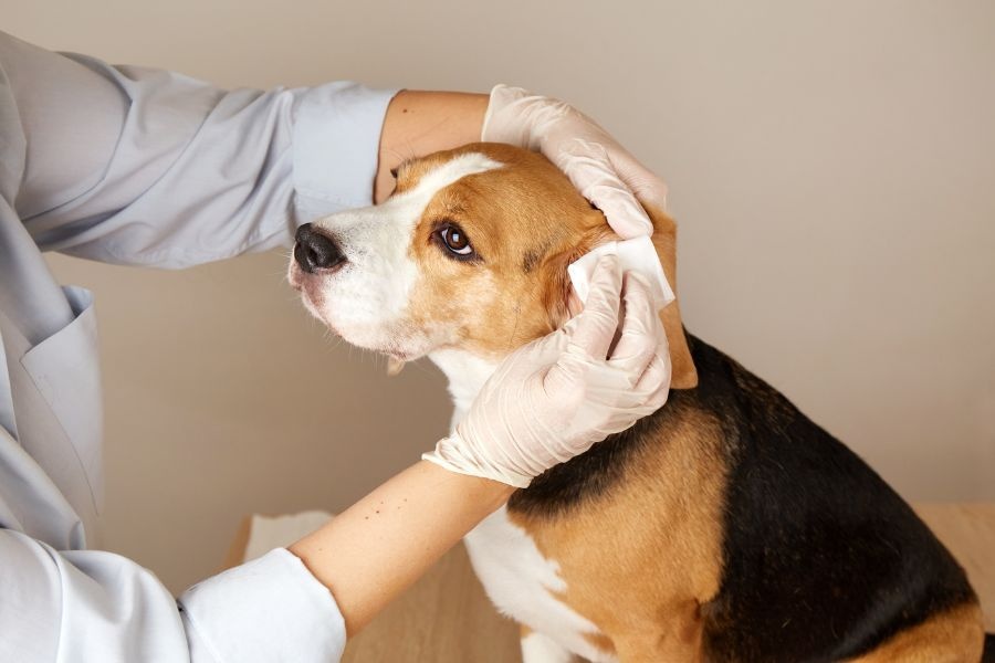
You might be here because something changed with your pet and you cannot quite name it. Maybe your dog started limping overnight, or your cat is losing weight even though bloodwork looks “normal.” You know something is wrong, yet you are staring at test results that do not add up. It is a lonely, anxious place to be when you are searching for a veterinary in Oakville, ON.
Then the veterinarian mentions X rays, an ultrasound, maybe even a CT or MRI. Your mind jumps straight to money, risk, and fear. You might wonder if your pet will be in pain, if the scan is really necessary, or if this is just “one more test.” That tension is real, and it deserves respect.
Here is the short version of what you need to know. Diagnostic imaging in veterinary medicine is not about doing more. It is about seeing what blood tests and physical exams cannot show, so your veterinarian can stop guessing and start treating with purpose. Used wisely, imaging can shorten the time to a diagnosis, avoid unnecessary surgeries, and give you a clearer picture of what your pet is facing.
So where does that leave you when someone is asking you to say yes or no to an imaging study for an animal you love?
Why does my vet recommend imaging if the exam seems “fine”?
One of the hardest parts of caring for animals is that they cannot point to where it hurts or explain what the pain feels like. A dog with a “simple” limp might have a torn ligament, a bone tumor, or a small fracture. A cat that hides more than usual might have arthritis, heart disease, or cancer. On the outside, they can look the same.
This is where veterinary diagnostic imaging starts to matter. X rays, ultrasound, CT, and MRI give a view inside the body that hands and eyes alone cannot match. They turn “I think” into “I can see.” Because of that, the choice is rarely about doing imaging for the sake of technology. It is about whether you are comfortable moving forward with treatment without knowing what is really going on.
Imagine these common situations.
You have an older large breed dog with a persistent cough. The physical exam is normal, and the heart sounds fine. Without chest X rays, your vet is guessing between infection, heart disease, or cancer. With X rays, they can see if there is fluid, masses, or an enlarged heart and target treatment instead of trying one medication after another.
Or you have a cat that stopped eating. Basic bloodwork looks okay. Without an abdominal ultrasound, you are left with broad guesses. With ultrasound, your vet might see thickened intestines that suggest inflammatory bowel disease, or a mass that requires surgery, or changes in the liver that call for a biopsy. The path forward becomes clearer.
So the real question becomes this. Are you comfortable treating in the dark, or do you want your medical decisions for your pet to be based on what is actually there?
What are the main imaging options for pets and what do they really show?
The phrase diagnostic imaging for animals covers several different tools, each with its own strengths and limits. Understanding them can help you feel more grounded when you hear these terms in the exam room.
For a visual overview of what different studies look like and which body parts they highlight, you can explore this helpful veterinary imaging anatomy resource.
Here are the most common imaging methods and what they are usually used for.
X rays (radiographs). These use a quick burst of radiation to create still images of bones and some soft tissues. They are often used for fractures, arthritis, hip and elbow issues, heart size, and basic chest or abdominal checks. Pets usually do not need sedation unless they are very painful or anxious.
Ultrasound. This uses sound waves, not radiation. It is excellent for looking at soft tissues like the liver, spleen, kidneys, bladder, intestines, and even the heart when done as an echocardiogram. It can also guide needle biopsies, which means smaller samples and fewer exploratory surgeries.
CT (computed tomography). CT scans create cross sectional images, a bit like slices through the body, and are especially helpful for complex bony areas like the skull, spine, nasal passages, and some chest or abdominal problems. They typically require sedation or anesthesia so the pet stays completely still.
MRI (magnetic resonance imaging). MRI is especially strong for brain, spinal cord, and some soft tissue problems. It is commonly recommended for seizures of unknown cause, suspected slipped discs, or some cancers. It always requires anesthesia, and it is usually done at specialty centers.
If you want a deeper technical explanation of how each of these works and when veterinarians use them, you can review this overview of veterinary diagnostic imaging.
Knowing what each tool is good at does not remove the worry, but it does give you language and clarity when you talk with your veterinarian.
How do the benefits and risks of imaging compare in real life?
Every test has tradeoffs. You are balancing the cost, the need for sedation or anesthesia, and any radiation exposure against the value of the information you gain. When you lay it out plainly, it becomes easier to decide what feels right for your pet and your family.
| Imaging Type | Typical Uses | Sedation / Anesthesia | Radiation | Common Benefits | Common Limits |
|---|---|---|---|---|---|
| X rays | Bones, chest, hips, basic abdomen | Rarely needed, except in very painful or anxious pets | Yes, low dose | Fast, widely available, relatively affordable | Soft tissues are not as clear, can miss early disease |
| Ultrasound | Abdomen, heart, soft tissues | Often no, sometimes mild sedation | No | Real time view, can guide biopsies, no radiation | Image quality depends on operator skill, limited for bones and lungs |
| CT scan | Head, spine, chest, complex fractures, some cancers | Usually yes | Yes, higher than X rays but still controlled | Very detailed images, great for planning surgery | Higher cost, requires advanced equipment and anesthesia |
| MRI | Brain, spinal cord, some soft tissue tumors | Always yes | No | Best soft tissue detail for the nervous system | High cost, limited availability, longer procedure time |
So what does this mean for you in a practical sense?
If your older dog has a new limp, starting with X rays often makes sense. They are fast and affordable, and they show fractures or advanced
well. If your cat has chronic vomiting and weight loss, ultrasound is often more informative than X rays, because it shows the layers of the intestines and the structure of the organs. If your young dog has sudden paralysis in the back legs, MRI may feel like a big step, yet it is often the only way to see the spinal cord clearly and decide if surgery could help.
When you look at it this way, imaging is less about choosing a fancy test and more about choosing the right level of detail for the problem in front of you.
What can you do right now to make better imaging decisions for your pet?
You do not need to become a medical expert to make good choices. You just need a clear process and a few key questions. Here are three steps you can take as soon as imaging comes up in conversation with your veterinarian.
1. Ask what your vet is trying to confirm or rule out
Before you agree to any study, ask your veterinarian to name the top two or three conditions they are worried about and how the imaging will change their plan. For example, “If the X rays show arthritis, we will manage pain. If they show a bone tumor, we will talk about surgery or palliative care.”
If the answer is vague, it is fair to say, “Can you walk me through how this diagnostic imaging will change what we do next for my pet?” A clear answer often makes the cost and effort feel more reasonable.
2. Talk openly about budget and timing
Money is part of real life. You are not being difficult if you need to understand costs up front. Ask for a written estimate that separates imaging, anesthesia or sedation, and any follow up like biopsies. Then ask if there are staged options.
For example, you might start with X rays and bloodwork, then move to ultrasound only if the first tests do not explain the problem. Or you might choose to do imaging at a referral center where all services are in one place, which can lower repeat visit costs.
Honest conversations about budget help your veterinarian tailor a plan that respects both your pet’s needs and your limits.
3. Prepare your pet and yourself for the day of imaging
Once you choose to move forward, small steps can reduce stress. Ask if your pet needs to be fasted. Bring familiar items like a blanket or toy if the clinic allows it. For anxious dogs or cats, ask about pre visit anxiety medication.
For studies that require anesthesia, ask who will monitor your pet, what safety protocols are in place, and when you can expect an update. Knowing the timeline and the plan helps you feel less helpless while you wait.
Finding peace with the choice to see “inside” your pet
Standing at this crossroads can feel heavy. You want to do everything, yet you also want to avoid putting your pet through more than they need. That tension is normal. It is a sign that you care deeply and that you are trying to be thoughtful, not reckless.
When used with intention, veterinary imaging services do not replace good clinical judgment. They sharpen it. They turn vague worry into clearer information, which can lead to kinder, more targeted treatment and, sometimes, the relief of knowing exactly what you are facing.
You do not have to rush your decision. Take a breath. Ask your questions. Make sure you understand what your veterinarian hopes to learn and how it will change the plan for your pet. Then choose the path that aligns with your values, your resources, and your pet’s comfort.
Your concern is already a powerful act of care. Whatever you decide, you are showing up for your animal, and that matters more than any single test.